
Life Science
PAXcam cameras are used in many life science applications where image detail and nuances of color are critical. Applications range from brightfield microscopy of stained histological slides, phase contrast microscopy for in vitro fertilization, fluorescence microscopy for cell biology studies, stereozoom microscopy for necropsy in pharmaceutical labs, and much more.
PAX-it software and its optional modules provide the imaging tools for easily cataloguing captured images and sharing them with colleagues. Stitching of adjacent fields of view for larger overviews, fusion of focal planes, blending of color channels, high dynamic range imaging, and many more image processing functions are part of the basic set of PAX-it software features. Reporting and sharing tools are easy to use; one click in PAX-it can send selected images to an Outlook email, Powerpoint presentation, or report in Excel or Word.
Optional tools are also available in additional software modules, to expand your capabilities even further. Measurements on images are easily accomplished, shown on the image and in a summary table, and exported directly to reports. Image analysis tools for life science applications include morphometric analysis, area fractions for color thresholding, optical profiles for otolith or “tree ring” analysis, and much more.
Motorized microscope controls and scripting tools are also available, to aid in the collection of images and data. Contact us for more information!
You may also be interested in:
- PAXcam cameras, for example for brightfield microscopy of stained histological slides, phase contrast microscopy for in vitro fertilization, fluorescence microscopy for cell biology studies, stereozoom microscopy for necropsy in pharmaceutical labs, and much more.
- Microscopes and macrostand hardware for your PAXcam camera: A variety of workstations may be integrated for imaging.
- Add-on software modules for basic measurements on images, or image analysis functions: measure features, or automatically detect objects or areas in your images.
- Motorized controls for automation of image collection and analysis
Life Science Images
Images taken with PAXcam cameras from various labs and samples.
NOTE: Images have been resized downward, and compressed, in order to present them as samples on the web. Original images are larger files and higher resolution.
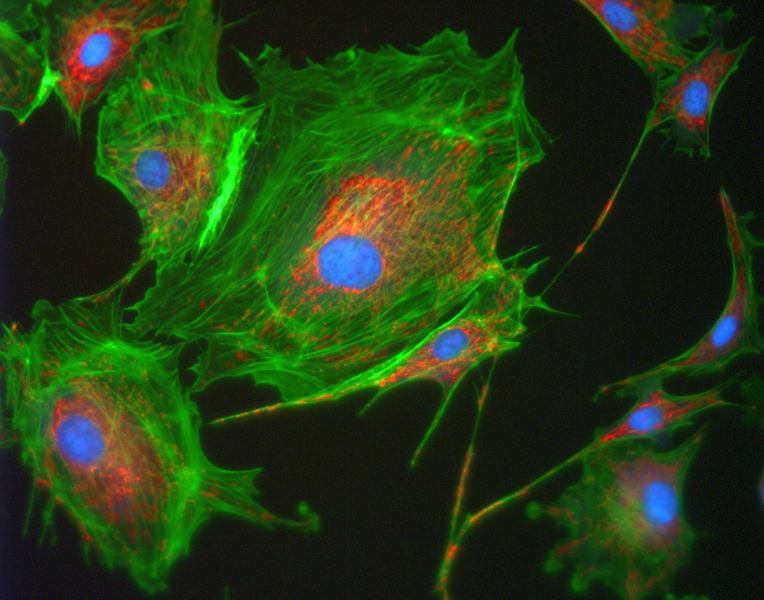
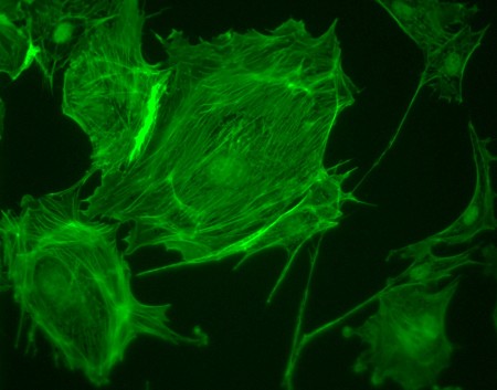
Images from R-G-B channels blended in PAX-it software

PAXcam2+; Sperm and epithelial cells, phase contrast and fluorescence images blended and annotated in PAX-it software
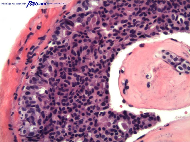
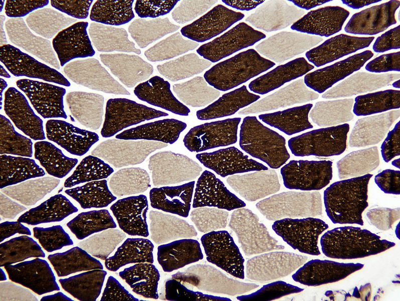
PAXcam3; Histo section with 20x objective
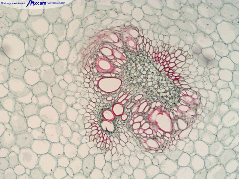
PAXcam2; Cross section of corn stem

PAXcam2; Lily anther long section
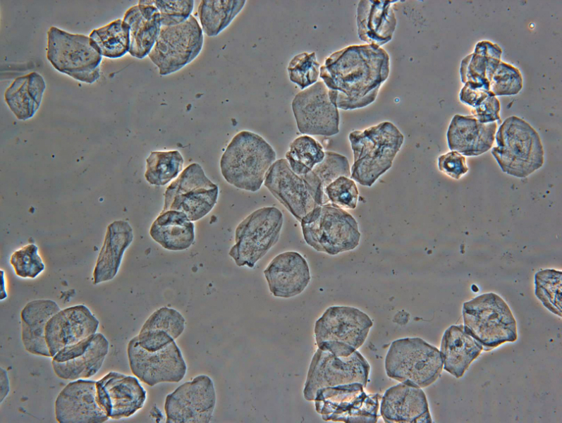
PAXcam2+; Epithelial cells, phase contrast
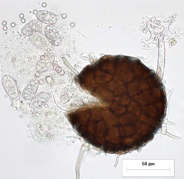
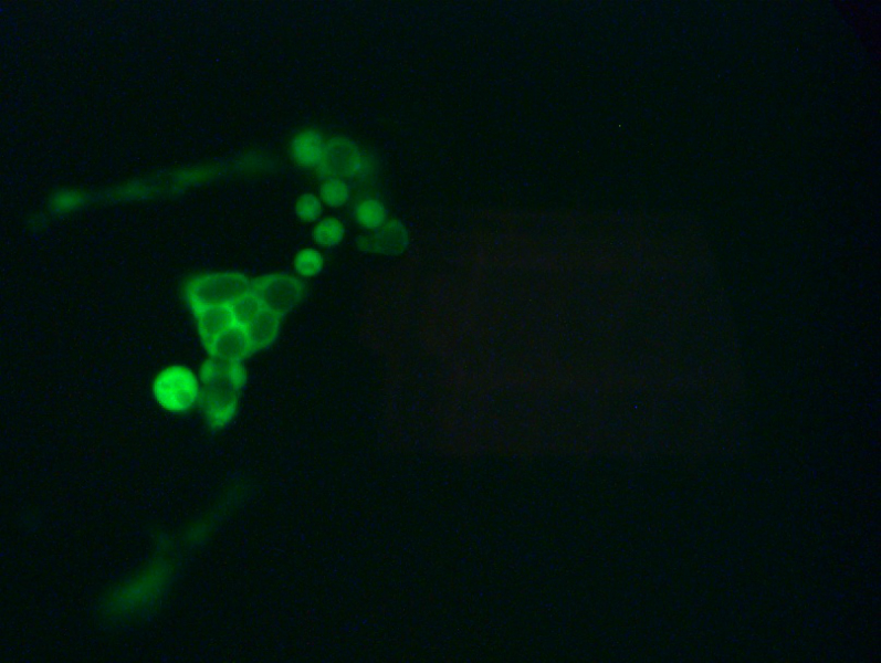
PAXcam ARC; Spore germination
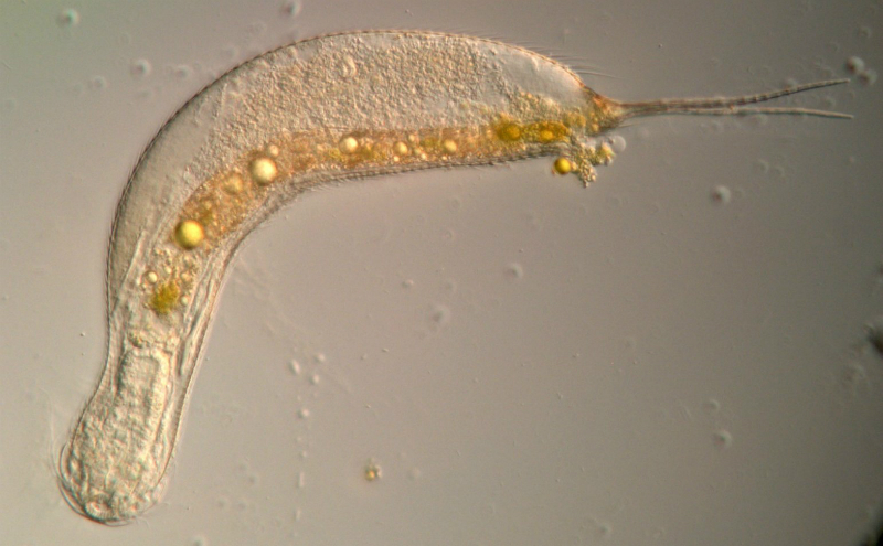
PAXcam2+, Gastrotrich P. nodicaudis, differential interference contrast (DIC).